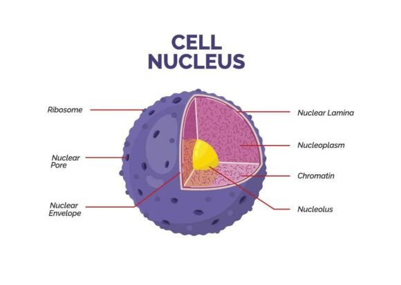
Labeled Diagram of Animal Cell Nucleus
If you're teaching biology, designing science presentations, or creating educational materials—even as a beginner—you’ll appreciate how much time and clarity a well-crafted Labeled Diagram of Animal Cell Nucleus saves. This isn’t just another clipart image. It’s a modern, flat-style biology education infographic—clean, accurate, and built for real-world use.
What Makes This Nucleus Diagram Different?
This diagram highlights the nucleus with precision: the nuclear envelope, nucleoplasm, nucleolus, and chromatin—all clearly labeled and proportionally balanced. Unlike generic illustrations, it emphasizes functional anatomy without overwhelming detail. Chromatin is shown as diffuse, thread-like material—not condensed chromosomes—so learners grasp its role in gene expression. The nucleolus appears as a distinct, dense region inside the nucleus, reinforcing its function in ribosome assembly.
It comes in three versatile formats: Vector (ideal for scaling without quality loss), EPS (compatible with professional design tools), and JPG (ready for quick web or slide use). Every element is 100% editable and resizable—no locked layers or flattened graphics. You can adjust colors in seconds, whether you’re matching a school’s brand palette, softening contrast for accessibility, or adapting visuals for print versus screen.
Why Educators and Creators Choose This Design
Teachers use it to replace blurry textbook scans or outdated PowerPoint diagrams. A high-school biology instructor might drop it into a Google Slides lesson on cell structure—then recolor the nucleolus in gold to highlight its role during protein synthesis. A homeschool parent could print it as a study poster, then annotate it by hand with their student. Freelance instructional designers integrate it into e-learning modules where interactivity matters: hover labels, zoomable sections, or animated transitions all start from this clean base.
Entrepreneurs building science-themed merchandise—like notebooks, flashcards, or classroom decor—rely on vector scalability. Need a tiny version for a sticker? A large banner for a science fair booth? Done. No pixelation, no redraws. Marketers promoting online biology courses use the JPG version directly in social media carousels—clear, on-brand, and instantly understandable.
Real Uses Beyond the Classroom
- Bloggers & Content Creators: Embed the diagram in an article about DNA replication or epigenetics—then add custom callouts using simple editing tools. Readers absorb complex ideas faster when structure is visual and labeled.
- Small Business Owners: Science tutors or edtech startups include it in client welcome kits or onboarding decks—adding credibility and reducing explanation time during first sessions.
- Freelancers & Designers: Use it as a foundation for custom infographics. Swap fonts, adjust spacing, or layer icons—all while preserving anatomical accuracy.
- Hobbyists & Lifelong Learners: Print it, laminate it, and keep it on your desk while reviewing AP Bio concepts—or sketch notes around it to deepen understanding.
What to Keep in Mind Before You Use It
While this Labeled Diagram of Animal Cell Nucleus is beginner-friendly, a few practical considerations help you get the most from it. First, check your software compatibility: Vector and EPS files work best in Adobe Illustrator, Affinity Designer, or Inkscape. If you only use Canva or Google Docs, the JPG version gives you flexibility—but remember that resizing beyond original dimensions may soften edges.
Color customization is easy, but thoughtful choices matter. Avoid low-contrast combos (e.g., light yellow chromatin on pale beige nucleoplasm) if sharing with students who have visual impairments. The included README file walks you through color replacement step-by-step—even if you’ve never opened a vector editor before.
Also, remember this is an animal cell nucleus diagram—so it intentionally omits a cell wall, chloroplasts, or large central vacuoles. That specificity helps avoid confusion when comparing plant and animal cells. If your project requires side-by-side comparison, this diagram pairs naturally with a companion plant cell graphic.
How It Fits Into Modern Biology Education
Today’s learners engage with science visually, quickly, and across devices. A static, cluttered diagram loses attention. This one uses flat design principles: minimal shading, consistent line weight, intuitive spacing, and clear typography. Labels don’t overlap. Arrows point unambiguously. Nothing distracts from the core idea—the nucleus as the control center, housing genetic material and coordinating cellular activity.
It supports active learning too. Because everything is editable, students can hide labels and test themselves. Educators can remove the nucleolus label and ask, “What structure is responsible for rRNA synthesis?”—then reveal it with a click. That kind of interaction builds deeper retention than passive viewing ever could.
A Final Note—and a Friendly Request
This Labeled Diagram of Animal Cell Nucleus was created with care for both scientific accuracy and everyday usability. Whether you’re explaining mitosis to a 10th grader, building a client-facing science presentation, or launching a biology podcast with custom show notes—it’s designed to work quietly, reliably, and effectively.
If you found it helpful, please take a moment to rate the design. Your feedback helps creators like me continue offering resources that are not only technically sound but truly supportive of how people learn and work today. Thank you for your support—it means a lot.